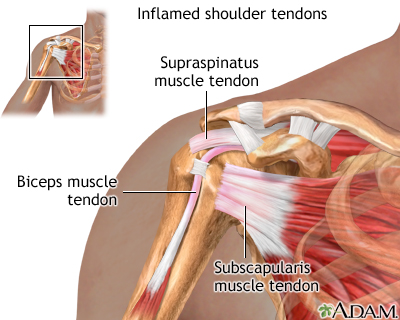
Shoulder Tendon Anatomy. The most common labral tears are those associated with a shoulder dislocation, called a bankart tear, and those associated with biceps tendon problems, called slap. It is the major joint connecting the upper limb to the trunk. The subacromial bursa lies on the superior aspect of the supraspinatus tendon (see the images below). • under normal conditions the amount of friction is reduced to a minimum by the large. The most common shoulder injuries involve the muscles, ligaments, cartilage, and tendons, rather than the bones.
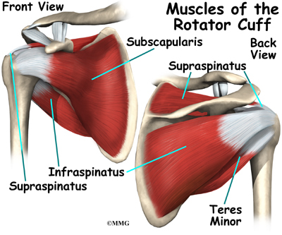
The rotator cuff tendons attach to the deep rotator cuff muscles. The shoulder is one of the largest and most complex joints in the body. • during abduction of the shoulder joint, the supraspinatus tendon is exposed to friction against the acromion. The subscapularis, teres minor, supraspinatus, and infraspinatus. The most common shoulder injuries involve the muscles, ligaments, cartilage, and tendons, rather than the bones.
Rotator Cuff Problems Medlineplus Medical Encyclopedia from medlineplus.gov The long head of biceps (lhb) is a very important tendon that travels through the shoulder joint (glenohumeral joint). Four muscles make up the rotator cuff: In addition to shoulder dislocations, other common injuries include rotator cuff tendon tears and broken bones including the humerus and collar bone. It has the greatest range of motion of any joint in the body allowing complete global movement allowing you to position the hand anywhere in space. Your shoulder is made up of three bones: The scapula (shoulder blade), clavicle (collarbone) and humerus (upper arm bone). The most common shoulder injuries involve the muscles, ligaments, cartilage, and tendons, rather than the bones. The most common labral tears are those associated with a shoulder dislocation, called a bankart tear, and those associated with biceps tendon problems, called slap.
Once the ligaments, tendons, and muscles around the shoulder become loose or torn, dislocations can occur repeatedly.
Ligation of the axillary artery. The most common shoulder injuries involve the muscles, ligaments, cartilage, and tendons, rather than the bones. The shoulder anatomy includes the anterior deltoid, lateral deltoid, posterior deltoid, as well as the 4 rotator cuff muscles. Your upper arm bone (humerus), your shoulder blade (scapula), and your collarbone (clavicle). There are actually four joints within the shoulder: Webmd's shoulder anatomy page provides an image of the parts of the shoulder and describes its function, shoulder problems, and more. Various types of injuries and degenerative conditions can cause the shoulder to become painful. Four muscles make up the rotator cuff: Sechrest, md narrates an animated tutorial on the basic anatomy of the shoulder. The shoulder joint is formed where the humerus (upper arm bone) fits into the scapula. Specifically, the four rotator cuff muscles include the following It has the greatest range of motion of any joint in the body allowing complete global movement allowing you to position the hand anywhere in space. Your shoulder is made up of three bones:
Shoulder anatomy is made up primarily of bones, muscles, ligaments, nerves and blood vessels. In this article, we shall look at the anatomy of the shoulder joint and its important clinical correlations. The rotator cuff tendons, and the biceps long head tendon are the most commonly injured. Shoulder radiology & anatomy at usuhs.mil. The arm bone (humerus), the shoulder blade (scapular) it's the long head that gives grief due to the long journey of its tendon through a groove in the humerus to its attachment just above the glenoid fossa.
Shoulder Anatomy Eorthopod Com from eorthopod.com This group of muscles lies just outside the shoulder joint. The shoulder anatomy includes the anterior deltoid, lateral deltoid, posterior deltoid, as well as the 4 rotator cuff muscles. • under normal conditions the amount of friction is reduced to a minimum by the large. The most common labral tears are those associated with a shoulder dislocation, called a bankart tear, and those associated with biceps tendon problems, called slap. The shoulder joint is formed where the humerus (upper arm bone) fits into the scapula. The rotator cuff tendons attach to the deep rotator cuff muscles. Rotator cuff and shoulder anatomy. The scapula (shoulder blade), clavicle (collarbone) and humerus (upper arm bone).
It has the greatest range of motion of any joint in the body allowing complete global movement allowing you to position the hand anywhere in space.
The long head of biceps (lhb) is a very important tendon that travels through the shoulder joint (glenohumeral joint). The rotator cuff tendons attach to the deep rotator cuff muscles. It is the major joint connecting the upper limb to the trunk. The scapula has several parts. The rotator cuff tendons, and the biceps long head tendon are the most commonly injured. Functional anatomy of the shoulder. An image depicting shoulder anatomy can be seen below. Once the ligaments, tendons, and muscles around the shoulder become loose or torn, dislocations can occur repeatedly. The subacromial bursa lies on the superior aspect of the supraspinatus tendon (see the images below). It has the greatest range of motion of any joint in the body allowing complete global movement allowing you to position the hand anywhere in space. Ligation of the axillary artery. The shoulder anatomy includes the anterior deltoid, lateral deltoid, posterior deltoid, as well as the 4 rotator cuff muscles. Chronic or acute wear and tear on the glenohumeral joint can lead to the painful tearing of the tendons of the rotator cuff or a torn labrum.
Three bones come together at the shoulder joint. It is the major joint connecting the upper limb to the trunk. • during abduction of the shoulder joint, the supraspinatus tendon is exposed to friction against the acromion. Your shoulder is made up of three bones: Understanding shoulder anatomy as well as the structures that are often injured can help in both prevention and treatment of sports injuries.
Rotator Cuff Problems Medlineplus Medical Encyclopedia from medlineplus.gov Related online courses on physioplus. Specifically, the four rotator cuff muscles include the following • during abduction of the shoulder joint, the supraspinatus tendon is exposed to friction against the acromion. Shoulder joint allows lifting, pushing and pulling by upper extremity. The long head of biceps (lhb) is a very important tendon that travels through the shoulder joint (glenohumeral joint). Sechrest, md narrates an animated tutorial on the basic anatomy of the shoulder. The scapula (shoulder blade), clavicle (collarbone) and humerus (upper arm bone). • under normal conditions the amount of friction is reduced to a minimum by the large.
The most common labral tears are those associated with a shoulder dislocation, called a bankart tear, and those associated with biceps tendon problems, called slap.
Chronic or acute wear and tear on the glenohumeral joint can lead to the painful tearing of the tendons of the rotator cuff or a torn labrum. Four muscles make up the rotator cuff: This group of muscles lies just outside the shoulder joint. The subacromial bursa lies on the top portion of the supraspinatus tendon. In the video below, dr. The subscapularis, teres minor, supraspinatus, and infraspinatus. Anatomy of the canine shoulder (scapula, humerus, ligaments, shoulder joint, muscles and tendons) on ct. There are actually four joints within the shoulder: Three bones come together at the shoulder joint. In this episode of eorthopodtv, orthopaedic surgeon randale c. The shoulder joint consists of 2 bones, the scapula and the humerus. Specifically, the four rotator cuff muscles include the following The shoulder is one of the largest and most complex joints in the body.